

Point-of-Care with Raman: Detection of cancer, infection, and more
The medical industry is a new frontier for Raman spectroscopy. In the past few years, Raman has been used in dental and cancer studies, and is now building on this success by extension into Point-of-Care (POC) applications. This article gives an overview of some very new and exciting research into the use of Raman spectroscopy for the detection of cancerous tissue, biomarkers of disease, and pathogens that cause disease.
Click on a link below to jump directly to a topic:
- Detecting bone infection with a handheld Raman spectrometer
- Raman spectroscopy for cancer detection
- Traditional Raman spectroscopy in breast cancer assessment
- SERS for detection and measurement of pancreatic cancer biomarkers
- Rapid, Point-of-Care (POC) assay for femtogram-level detection of COVID-19
- Multiplex immunophenotyping of blood and breast cancer cells with Raman spectroscopy
- Easy detection of enzymes with the electrochemical-SERS effect
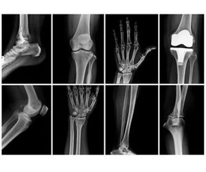
Complication from infection is a serious issue for the use of human bone grafts in musculoskeletal surgery.Staphylococcus epidermidisandStaphylococcus aureusare the most common culprits in bone-related infections, and treatment of these bacteria is extremely difficult.
Detection of staph bacteria on graft material and distinction between healthy and diseased bone are both crucial in preventing infection. Laboratory culture traditionally requires 7–10 days for results and carries the risk of contamination during transport and testing. In positive cases, the patient must be treated retroactively with massive doses of antibiotics. An ideal solution for this challenge is on-site analysis that permits the surgical team to identify and avoid diseased bone on the spot.
Recently, a research group in Austria demonstrated successful differentiation between healthy and infected bone samples and between two types of staph bacteria using a handheld MIRA Raman spectrometer [1]. Their procedure analyzed the fingerprint Raman bands of phosphates, amides, and collagens, and their differing intensity and peak width ratios to distinguish between healthy and diseased bone. Principal component analysis (PCA) supported optical analysis and was used in the finer distinction of staph strains.
The Austrian group appreciated that MIRA was «lightweight, compact, and battery powered» and that Raman spectroscopy has the benefit of «minimal sample preparation and rapid results.» Testing required only a tiny bone sample for in-situ examination during surgery and provided fast and accurate results directly in the operating room.
Raman’s «molecular fingerprint» spectra can be sensitive enough to detect the chemical changes that accompany disease. Compact Raman spectrometers can help surgeons assess tumors during surgical procedures for faster decision-making.Application examples for breast cancer and pancreatic cancer are discussed in the following sections.

When breast cancer is suspected, one surgery is typically required for biopsy and a second is for removal of malignant tumors. The ability to assess suspect tissues during preliminary surgery permits immediate removal, if required. The benefit to the patient and to the medical industry is inestimable—and research suggests that Raman spectroscopy may be able meet this need for some types of cancer.
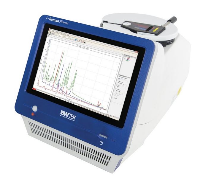
Raman spectroscopy is sensitive enough to detect changes in tissue that result from many types of cancer. For example, there are very subtle differences in Raman spectra from healthy breast tissue and malignant tumors. Researchers in the UK used a high-resolution i-Raman laboratory device (Figure 1) and multivariate tools, including PCA, for successful discrimination of healthy and cancerous tissues [2].
SERS (surface-enhanced Raman spectroscopy) can be a solution when Raman is not ideal for analysis. This may be the case in situations where the target exists in a complex sample matrix, or because of the fluorescence of carbon-based molecules. SERS enhances the Raman signal, but not the competing fluorescence signal. Also, the SERS effect enables sensitive detection of analytes at mg/L levels—sometimes down to µg/L. Finally, SERS peaks are sharp and well-defined, allowing efficient detection and identification of target analytes.
To learn more about SERS, read our previous blog post on this topic.
Raman vs SERS… What’s the Difference?
Pancreatic cancer is deadly, partly because it is hard to diagnose. However, there are some biomarkers that are present at elevated levels in ~75% of positive cases [3]. These can be detected with enzyme-linked immunosorbent assays (ELISA), which measure a variety of biomarkers, including antibodies, antigens, and proteins.
In an emerging technique, an i-Raman laboratory spectrometer was used at the University of Utah for SERS analysis in conjunction with ELISA to detect an antigen associated with pancreatic cancer [2]. The SERS signal was generated from a reporter molecule complexed to both a gold nanoparticle and the target analyte in an otherwise classical lateral-flow or «sandwich» immunoassay (Figure 2). This is an incredibly accurate technique, enabling very sensitive detection – with potential for quantification – of the biomarker of interest.

Rapid, Point-of-Care (POC) assay for femtogram-level detection of COVID-19
\r\n\r\n
Lateral flow immunoassays provide relatively quick results, but they deliver nanogram-level detection and have quantification limitations. In comparison, the SERS-based ELISA is sensitive tofemtogram quantitiesof antigen, with fast results at the POC with a commercial handheld Raman instrument.
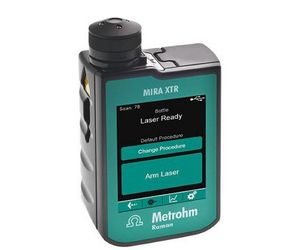
An alternate ELISA format was used by researchers at the University of Wyoming to detect antigen biomarkers associated with COVID-19 infection [4]. This study employed a magnetic nanoparticle-supported assay to concentrate the target biomarker in solution for subsequent SERS detection with MIRA XTR (Figure 3). It proved to be more sensitive than commercial lateral flow assays, was compatible with both solvent and saliva samples, could be adapted to new virus variants, and achieved highly sensitive POC diagnosis of COVID-19.
Lateral flow immunoassays provide relatively quick results, but they deliver nanogram-level detection and have quantification limitations. In comparison, the SERS-based ELISA is sensitive tofemtogram quantitiesof antigen, with fast results at the POC with a commercial handheld Raman instrument.
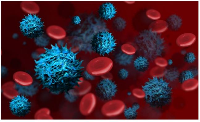
Another study used MIRA DS to evaluate a portable SERS-based ELISA for immunophenotyping of different types of red blood and breast cancer cell surfaces [5]. Distinguishing healthy and diseased cells and detection of multiple biotargets in one sample can support informed treatment of different kinds of breast cancer.
This assay had «specificity, sensitivity, and repeatability for… immunophenotyping in different cell types [using] smaller analysis sample volume as compared to conventional... multiplex immunoassays,» and it was less «labor-intensive and technically straightforward to perform.» The authors praised MIRA’s Orbital Raster Scan for improving the sensitivity of their assay through a larger area of interrogation and spatially averaged measurements.
Traditional multiplexing flow assays can be limited by availability of different colored stains and interpretation of results. They are also associated with a large laboratory footprint. In contrast, this method based on handheld Raman spectroscopy offers the potential for accurate POC results with quick sample-to-result time, multiplexing capability, and a very compact instrument
Easy detection of enzymes with the electrochemical-SERS effect
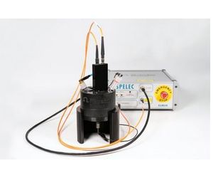
A very different SERS technique for characterizing biological molecules has been reported by Metrohm [6]. Electrochemical SERS (EC-SERS) facilitates two experiments at once: electrochemical activation of SERS features of silver electrodes (Ag SPEs) followed by spectroscopic detection of the sample (with SPELECRAMAN638, Figure 4).
Here, the SERS substrate is generated in-situ from silver electrodes (screen-printed as well as conventional). This is accomplished in the presence of the analyte while under continuous interrogation with Raman to optimize detection of SERS-active species. The 638 nm excitation provides a SERS effect with good intensity, with less risk of sample damage and fluorescence.
Determining the structure of enzymes (and their role in disease) such as aldehyde dehydrogenase (ALDH) helps to understand diseases. With EC-SERS, application scientists defined previously unreported fingerprint Raman bands of ALDH in solution. Similarly, the redox states of cytochrome c provide information about electron transport across cell membranes [7]. Cytochrome c changes oxidation- and conformation-state during the EC experiment, and these redox states possess distinguishable SERS spectra.
Raman technology is truly being utilized in innovative ways. Research groups worldwide are applying its considerable benefits – including sensitivity, trace detection, small form-factor, and fast results – for detecting cancerous tissue, disease biomarkers, and disease-causing pathogens. The findings are incredibly exciting and promising!