

Aiforia Technologies software
Aiforia Clinical Suites - CE-IVD Marked Clinical AI Models
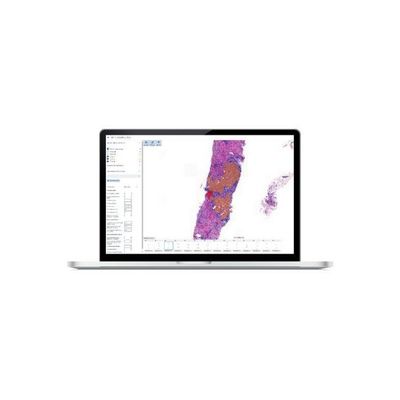
Aiforia - Clinical AI Model for Prostate Cancer, Gleason Grade Groups
Rapidly distinguishes between normal and cancerous tissue supporting the pathologist in the quick detection of tumor areas. Automatically scores a quantitative Gleason grade score and grade group from WSI, saving the pathologist time. Provides visual feedback as it allows to view images in different magnifications, move in different x-y-z-locations, view image analysis results, and mark and measure significant features in the images.
Aiforia - Model PR - Clinical AI Model for Breast Cancer
Creates a heatmap assisting the pathologist in automatically finding the critical areas of the sample. Rapidly distinguishes between normal and cancerous tissue supporting the pathologist in the quick detection of tumor epithelium. Automatically scores the PR positive and negative cells of epithelial origin from either WSI or selected image areas, saving the pathologist time. Provides intelligent viewing as the PR AI Model enables viewing and selection of areas with high density of PR-positive cells, or hotspots, displaying the results of image analysis.
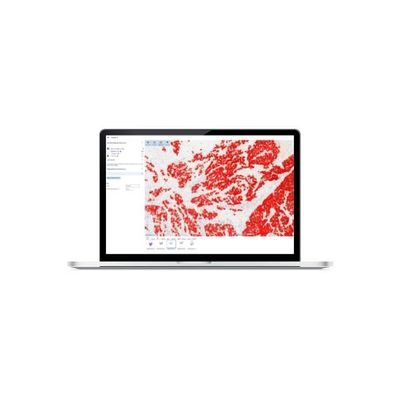
Aiforia - Model ER - Clinical AI Model for Breast Cancer
Creates a heatmap assisting the pathologist in automatically finding the critical areas of the sample. Rapidly distinguishes between normal and cancerous tissue supporting the pathologist in the quick detection of tumor epithelium. Automatically scores the ER positive and negative cells of epithelial origin from either WSI or selected image areas, saving the pathologist time. Provides intelligent viewing as the ER AI Model enables viewing and selection of areas with high density of ER-positive cells, or hotspots, displaying the results of image analysis.
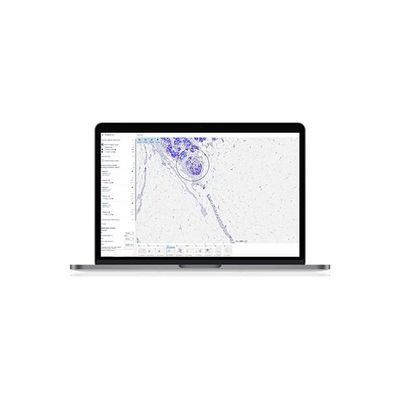
Aiforia - Model Ki67 - Clinical AI Model for Breast Cancer
Creates a heatmap assisting the pathologist in automatically finding the critical areas of the sample. Rapidly distinguishes between normal and cancerous tissue supporting the pathologist in the quick detection of tumor epithelium. utomatically scores the Ki67 positive and negative cells of epithelial origin from either WSI or selected image areas, saving the pathologist time. Provides intelligent viewing as the Ki67 AI Model enables viewing and selection of areas with high density of Ki67-positive cells, or hotspots, displaying the results of image analysis.
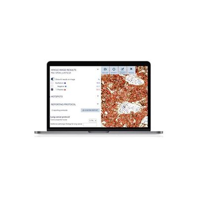
Aiforia - Model PD-L1 - Clinical AI Model for Lung Cancer
Creates a heatmap assisting the pathologist in automatically finding the critical areas of the sample. Rapidly distinguishes between normal and cancerous tissue supporting the pathologist in the quick detection of tumor epithelium. Automatically scores the PD-L1 positive and negative cells of epithelial origin from whole slide images (WSI) saving the pathologist time when evaluating patient samples. Provides intelligent viewing and selection of areas with high density of PD-L1 positive cells displaying the results of image analysis in different magnifications and enables the user to move in different x-y-z-locations, as well as to mark and measure features in the images.
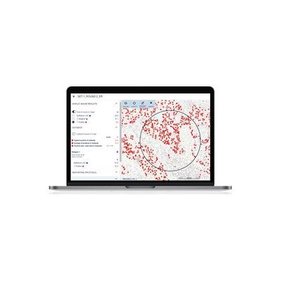
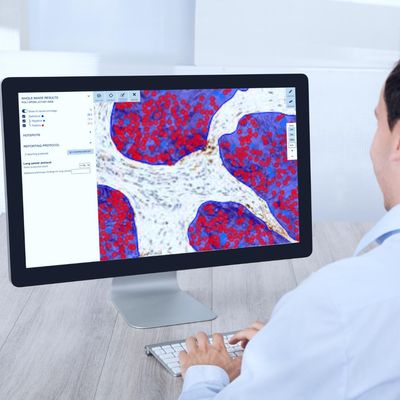
Aiforia - Clinical Suite Viewer
Rapidly displays AI-assisted analysis results in an intuitive and easily readable format and enabling case prioritization based on severity. Automatically generates reports from analyses by clinical AI models, significantly saving the pathologist time and improving the lab’s overall output efficiency. Supports full interaction with the pathologist by allowing viewing of images in different magnifications, movement in different x-y-z-locations, viewing of image analysis results in high precision, and markings and measurement of significant features in the images. Users can easily edit the results in the report, select hotspot areas and exclude control tissue or areas of bad quality.
Aiforia - Platform for QC and Adaptation
An intuitive user interface in which you can easily adapt your AI model to accommodate changes if they are needed. Easy management and monitoring for QC and auditing purposes. Compliant with the FDA’s Good Machine Learning Practices (GMLP), a framework outlining best practices in software engineering and quality management of AI systems.
Research
Aiforia - Create for Cloud-Based AI Software for Image Analysis
Automate manual tasks, standardize analysis, and find the hard to spot objects with our cloud-based AI software for image analysis. Aiforia Create is made for seamless use by the medical professional to create and use AI models to increase the speed and accuracy of any image analysis application.