

- Home
- Companies
- Envision Radiology
- Services
Envision Radiology services
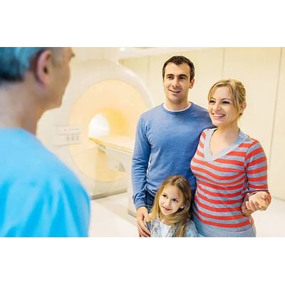
Arthrogram MRI Services
Joints are essential parts of your body that give you freedom of movement, but unfortunately, they can go wrong in many ways. An arthrogram MRI allows radiologists to pinpoint issues in your joints that standard imaging may miss. Arthrogram, also called arthrography, is a series of images taken using an X-ray, MRI, CAT scan or fluoroscopy. Before the arthrogram MRI procedure, your joint is injected with a contrast dye, usually iodine. Using fluoroscopy, the radiologist guides the placement of the injection into the joint. The contrast dye coats the inner lining of the joint structures which makes it appear white on the arthrogram and helps highlight what’s gone wrong in your joint. Using the images, your physician can assess the function of the joint, make a diagnosis and come up with a treatment plan.
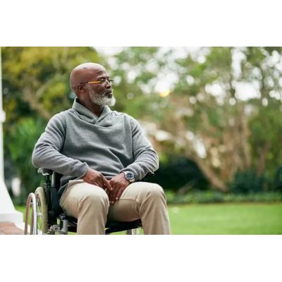
Bone Density Scan Services
Bone density scans also referred to as bone mineral density testing, is a medical test used as an indirect indicator of osteoporosis. This disorder makes your bones more fragile and likely to break. When you undergo a bone density scan, it measures the amount of mineral material per square centimeter in your bones. The bone density test can show if your bones are at risk of fracturing by using X-rays to measure the grams of calcium and bone minerals packed into a segment of bone. Generally, radiologists will test the spine, the hip or the forearm. High bone mineral content indicates denser bones. The denser your bones are, the stronger they are and less prone to breakage.
Cardiac Scoring Services
If your doctor needs to determine the amount of calcium or plaque in your coronary arteries, they may recommend getting cardiac scoring. This procedure takes less than 10 minutes of your time and is completely non-invasive and pain-free. Computed tomography, more commonly referred to as CAT scan technology, is used to take 70 to 90 images of your coronary arteries to establish your cardiac score.
CAT/CT Scans Services
A CAT Scan or computed tomography (CT), is a medical imaging method that produces a volume of data that can be manipulated (through a process known as windowing) to demonstrate various bodily structures based on their ability to block the x-ray beam. A CAT is created by combining a series of x-ray views taken from different angles. Modern scanners allow for 3-D representations of structures. CT scans show bones and soft tissues inside the body. Medical professionals can view the images individually or as an entire view in 3D. This type of technology is extremely valuable to doctors needing to make decisions very quickly.
CTA (CT Angiography) Services
A computed tomography angiography or CT angiography is a medical test in which a patient will receive an injection of a special dye through an IV in their arm or hand before undergoing a CT scan to produce detailed pictures of blood vessels and tissues. Computerized tomography scans or CT scans are X-rays that create cross-sectional images of your body with a computer. The CT scanner consists of a machine with a tunnel. During a CT angiogram, a patient will lie on an examining table and pass in and out of the tunnel while the machine scans a specific part of their body. Combining a CT scan with the contrast material dye causes blood vessels and tissues a clinician wishes to study further to ‘light up’ for further examination.
DEXA Scans Services
Physicians perform DEXA scans for a variety of reasons. The exam is most commonly used to diagnose osteoporosis and other conditions that cause bone loss. Physicians recommend DEXA scans for patients who fit into any of the following categories: Post-menopausal women who are not taking estrogen, Individuals with personal or maternal histories of smoking or hip fractures, Men with clinical conditions associated with bone loss, including chronic kidney or liver disease and rheumatoid arthritis, Patients who have experienced a fracture or mild trauma, Patients who take medications known to lead to bone loss, Individuals who have high bone turnover, Patients who have thyroid or parathyroid conditions.
Enterography Imaging Services
Enterography is an imaging study that produces high-resolution images of the small intestine and abdominal organs. It is a noninvasive procedure that is valuable for the diagnosis of inflammation, bleeding, obstructions and other gastrointestinal problems.
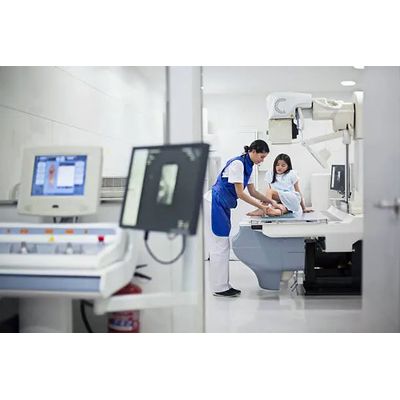
X-Ray Fluoroscopy Services
Fluoroscopy is an imaging technique that allows physicians to look at moving bodily structures in real-time. Many compare this to an X-ray movie because images are transmitted to a TV-like monitor so the body parts’ motion can be seen in detail. A fluoroscope utilizes an X-ray source and a fluorescent screen. The X-ray beam passes through the body part being examined. Some of the most common bodily structures analyzed using fluoroscopy include bones, muscles, joints, and organs like the lungs, heart, kidneys and more. Using the images created, physicians can look at many body systems in great detail.
IVP Protocol Services
CT Intravenous Pyelogram (CT IVP or CT Urogram) is an exam that uses an injection of contrast material into the veins followed by Computed Tomography imaging to evaluate kidneys, ureters, and bladder. The exam helps to diagnose urinary tract disorders such as kidney stones, urinary tract obstruction, or cancer. Often symptoms for this procedure are, but not limited to, hematuria (blood in the urine), flank (back) pain and abnormal lab work. When contrast material is injected into a vein in the patient’s arm, it travels through the bloodstream and collects in the kidneys and urinary tract, turning these areas bright white on the x-ray images. A CT IVP allows the radiologist to view and assess the anatomy and function of the kidneys, ureters and the bladder and is a valuable tool for diagnosis and treatment.
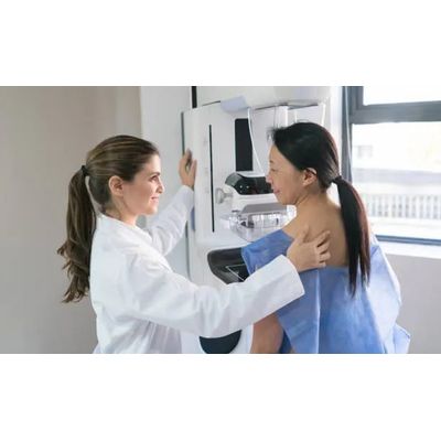
Mammography Services
A mammogram procedure is simply an X-ray image of your breast. Mammography uses a low dose X-ray to look for early signs of breast cancer, such as lumps that are too small to be felt either by yourself or your healthcare provider. A mammogram can also show changes in your breast tissue which could indicate early-stage breast cancer. Our radiologists use digital mammogram imaging to both locate and diagnose cancer nodules which were undetectable on older systems. This makes regular mammograms the best way to find breast cancer early on, sometimes as much as three years before it can be felt.