

Newcells Biotech Limited products
Lung Airway Models

Human Lung Fibroblasts
Human lung fibroblast model to test the efficacy of anti-fibrotic agents in vitro. Newcells human primary lung fibroblast model is the basis of our FMT assay to evaluate anti-fibrotic therapies in vitro at high throughput. The assay replicates extracellular matrix deposition (ECM) observed in vivo when lung fibroblast injury leads to activation and proliferation. Following injury, pulmonary fibroblasts proliferate playing an important role in the repair and remodelling of tissue. However, excessive proliferation of fibroblasts can result in abnormal tissue function.
Models of the Nephron

Glomerulus
Podocyte model for assessing glomerular toxicity. Newcells now offers the first fully differentiated, primary podocyte model derived from fresh kidney tissue for the in vitro assessment of drug-induced glomerular toxicity. The effect of a drug on the glomerular filtration barrier and podocyte glomerular permeability can now be easily evaluated in vitro. Podocyte injury and proteinuria can be modelled in vitro at high throughput (96-well format) and assessed by measuring podocyte damage biomarkers, TEER and podocyte permeability. Podocytes maintain the glomerular filtration barrier and similarly to proximal tubule cells, can be damaged by drugs. Drug-induced glomerular toxicity occurs progressively. First podocyte injury leads to cellular dedifferentiation causing perturbation of the podocyte monolayers architecture.

Proximal Tubule
Kidney proximal tubule cell model for safety/efficacy and transporter studies. aProximate™ is one of the most advanced, near physiological, in vitro, kidney proximal tubule cell (PTC) models. aProximate™ PTCs are derived from fresh human kidney tissue and grown on Transwells® where they remain as a functional polarised cell layer that forms tight junctions. In contrast to other primary and immortalised kidney proximal tubule cells in culture, aProximate™ PTCs retain high expression of the key transporters involved in drug handling including Megalin and Cubilin, which is ideal for drug transporter studies. Studies with aProximate™ can give you a detailed mechanistic understanding of how new drugs are transported and eliminated through the kidney and how they interact with other drugs prescribed to the target patient population to help mitigate risk of renal toxicity.
Retina Models
Retinal Organoids
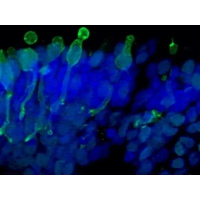
Light responsive retinal organoids for accurate prediction of clinical outcomes. The retinal organoids recapitulate the complex structure of the human retina with laminar cell organisation mimicking embryonic development. They contain the outer photoreceptor segment of the retina that responds to light. Available analytical readouts: Immunofluorescence analyses, mRNA quantification by RT-qPCR, Transcriptomic analysis by single-cell RNA sequencing, Cytotoxicity assays, Cytokine release, Flow cytometry, Electron microscopy
Retinal Pigment Epithelium (RPE)
A functional monolayer in vitro model of retinal pigment epithelial cells generated from human iPSCs for accurate prediction of clinical outcomes. The retinal pigment epithelial (RPE) cell model is composed of a monolayer of RPE cells cultured in 24-well Transwell® plates. RPE characterisation includes: morphology assessment, pigmentation, RPE-specific expression at the protein level (BEST1, TYRP1), the analysis of phagocytosis of photoreceptor outer segments, trans-epithelial resistance (TEER), polarity of apical Pigment Epithelium-Derived Factors (PEDF) and basal vascular endothelial growth factor (VEGF) secretion.