

Virasoft Inc. products
Virasoft - Telepathology Mobile System
The Telepathology Mobile System includes an apparatus that connects the microscope and your personal phone, and a mobile and web application that enables you to manage your consultation processes. In this way, you can consult your microscopic cases with your phone and save them in the system without the need for expensive scanning devices.
Decision Support & AI
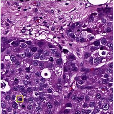
Virasoft - Mitosis Analysis Technology
In reporting of breast cancer pathology, mitosis detection and counting of mitotic cells are important factors that affect the tumor grade and the treatment of the patient. Mitotic cell counting requires standardization to a fixed field area. The total number of mitoses per 10 HPF is recorded. Normally, the count is performed manually by pathologists, but automating the process could reduce its time, minimize errors, and improve the comparability of results obtained in different pathology laboratories . Mitosis detection is a complex process because only definite mitotic figures have to be counted; hyperchromatic and pyknotic nuclei are ignored since they are more likely to represent apoptosis rather than cells in mitosis [1]. Therefore, an artificial intelligence method, convolutional neural networks (CNN), is used for mitosis detection in this analysis module.
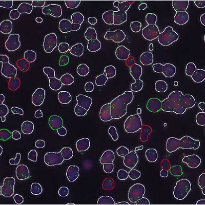
Fluorescent - Fluorescent in Situ Hybridization Technology
Fluorescent in situ hybridization (FISH) is a molecular technique commonly used to detect chromosomal abnormalities and gene mutations such as translocation, amplification, deletion. The desired gene regions are made visible with fluorescent labeled DNA probes, which is the complement of the gene region that needs to be detected [1]. Automatic FISH analysis detects amplification, break apart, fusion, deletion of target sequence and performs quantitative analysis in digital slides. It enables detection of very low signal and visualizes the DAPI and signals. The algorithm categorizes the nuclei to normal, abnormal and artifact groups.
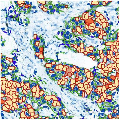
Virasoft - Model HER2 - Epidermal Growth Factor Receptor 2
The HER2 gene is an oncogene encoding the “Epidermal Growth Factor Receptor 2” which is a transmembrane glycoprotein. Amplification of the HER2 gene and overexpression of HER2 receptor on the cell membrane is observed in approximately %15 of invasive breast cancer.
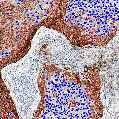
Virasoft - Model PD-L1 - Programmed Cell Death Protein
Programmed cell death protein 1 (PD-1; also called CD279) is one of the co-inhibitory receptors that is expressed on the surface of antigen-stimulated T cells [1]. Normally, PD-L1 expression can be detected on hematopoietic cells including T cells, B cells, macrophages, dendritic cells, mast cells and healthy tissue cells. But PD-L1 can be expressed by tumor cells and tumor stroma. Interaction tumoral PD-L1 with PD-1 stimulates PD-1-mediated T cell inhibition and down regulation of immune response. PD-L1-PD-1 pathway has proven value as a therapeutic target in a large number of malignancies including non-small cell lung cancer [2]. Immunohistochemical staining is a standardized approach to PD-L1 expression in order to distinguish for patients who may benefit from therapies [3]. Virapath PD-L1 analysis provides accurate quantification of positive cell proportion