

ProImmune - Human MR1 Tetramers
Human MR1 Tetramers detect MAIT cells with human MR1 tetramers. ProImmune is the leading commercial source worldwide for fluorescently labeled human MR1 tetramers. MR1 complexes bind to T cell receptors on mucosal-associated invariant T (MAIT) cells. MAIT cells have a particular specificity (as determined by the relevant MR1-binding small molecule ligand used), allowing identification, enumeration and isolation of antigen-specific MAIT cells by flow cytometry. Our tetramers show excellent brightness and experimental reproducibility. To avoid the difficulties associated with dissolving and loading the most commonly used ligands, 5-OP-RU and 6-FP, our tetramers come pre-loaded (not available in Australia and the United States). They can also be supplied empty for loading with your ligand of choice.
- empty ready for loading
- pre-loaded with 5-OP-RU
- pre-loaded with 6-FP (negative control)
- R-PE or APC labeled
- 50 or 150 tests
- Delivery in 1-2 days
- Guaranteed for 6 months
T cells can discriminate between foreign and host molecules by recognizing not only peptide and lipid antigens, but also riboflavin precursors found in many bacteria and yeasts. Presentation of these molecules on MR1 can selectively activate mucosal-associated invariant T (MAIT) cells. MAIT cells form an abundant population of innate-like T cells. MR1-presented MAIT cell activating antigens include 5-(2-oxoethylideneamino)-6-d-ribitylaminouracil (5-OE-RU) and 5-(2-oxopropylideneamino)-6-d-ribitylaminouracil (5-OP-RU). These are formed by non-enzymatic reactions between 5-amino-6-d-ribitylaminouracil (5-A-RU), an early intermediate in bacterial riboflavin synthesis, and glyoxal and methylglyoxal. MR1 binding adducts, such as 5-OP-RU are part of the microbial derived molecular signature of MAIT cell immunoreactivity.
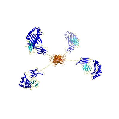
Human MR1 Tetramer
Human MR1 tetramer pre-loaded with 5-OP-RU compared to human MR1 tetramer pre-loaded with 6-FP (negative control), APC labeled, and stained on human PBMCs.
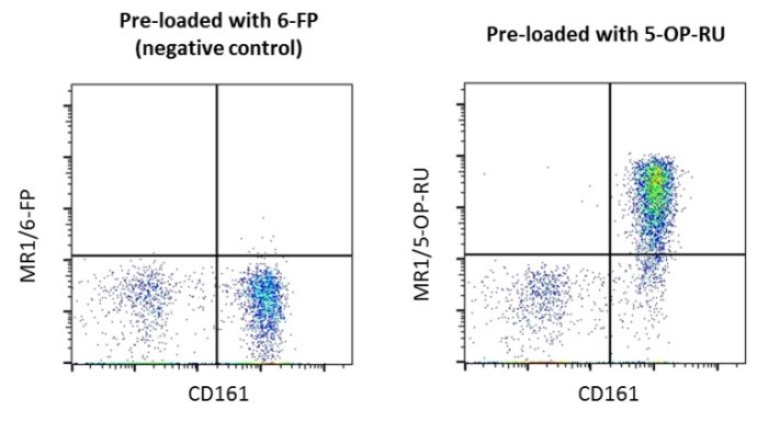
Figure 1. Human PBMCs stained with Negative Control (6-FP) and 5-OP-RU-loaded MR1 tetramers. A population of MAIT cells is clearly visible in the upper right quadrant following staining with MR1/5-OP-RU tetramer. MAIT cells were identified by first gating on CD19–/CD3+/TCR Vα7.2+ cells before plotting CD161 vs MR1 tetramer.