

Topcon Healthcare, Inc.
- Home
- Companies
- Topcon Healthcare, Inc.
- Products
- Topcon - Model Triton - Multimodal ...
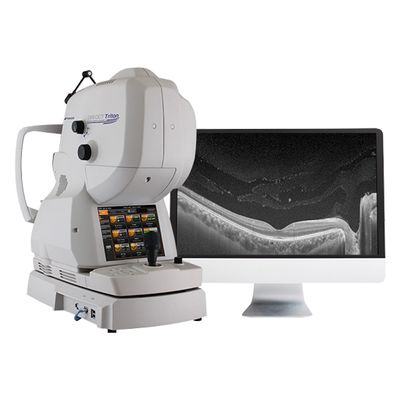
Topcon - Model Triton -Multimodal Swept-Source OCT Technology
Triton uses Swept-Source technology to allow visualization into the deepest layers of the eye – even through cataracts, hemorrhages, gas bubbles, and other media opacities, making it possible for more patients to be imaged.
The fast 100 kHz scanning speed and invisible scan beam rapidly capture detailed images, resulting in fewer motion artifacts and stunning image quality. Decrease chair time and improve your clinical workflow with fewer rescans and multimodal imaging including, SS-OCT, True-Color, Digital Red-free, and optional Anterior Segment OCT. Triton (plus) adds fluorescein angiography (FA) and fundus autofluorescence (FAF) to the Triton.
Most popular related searches
- Deep penetration through media opacities such as cataracts and hemorrhages
- Multimodal Imaging: SS-OCT + Non-mydriatic true color fundus imaging, FA and FAF available²
- Stunningly detailed images with 100k A-scans/sec. and 1,050nm wavelength
- Invisible scan beam allows patients to focus on the fixation target and reduce involuntary eye movement
- HIGH RESOLUTION: Multimodal platform provides easy, yet comprehensive comparison of microvascular impairment with FA1, FAF1, OCT and color fundus images.
- SCAN MORE PATIENTS: Swept-source technology allows imaging through media opacities.
- FEWER RESCANS: Invisible scan beam allows patients to focus on the fixation target and reduce involuntary eye movement.
- WIDE SCAN: A 12mm x 9mm scan encompasses the optic nerve and macula and can be acquired in 1.8 seconds for fast assessment of the posterior pole.
- RICH, DETAILED IMAGES: Image quality is further enhanced by PixelSmart® Technology2
Fundus Imaging
- Field of View : 45° / 30° (Digital Zoom)
- Operating Distance : 34.8mm
- Minimum Pupil Diameter : Ø4.0mm / Small Pupil Mode: Ø3.3mm
- Resolution (On Fundus) : Center: 60 Lines/mm or more,
- Middle (r/2): 40 Lines/mm or more,
- Periphery (r): 25 Lines/mm or more
OCT
- Scan Range (On Fundus) : 6 to 12mm
- Scan Patterns : 3D Wide: 12x9mm
- 3D Macula: 7x7mm
- 3D Optic Disc: 6x6mm
- Combination Scan: 12x9mm + 5 Line Cross
- Line: 6-12mm
- 5 Line Cross: 6-12mm
- Scan Speed : 100,000 A-Scans Per Second
- Lateral Resolution : 20 μm
- Axial Resolution : Optical: 8 μm
- Digital: 2.6 μm
- Minimum Pupil Diameter : Ø2.5mm
- Fixation Target : Internal Fixation Target/ Peripheral Fixation Target / External Fixation Target
- Diopter Range : Without the diopter compensation lens: -13D to +12D
- When the concave compensation lens is used: -12D to -33D
- When the convex compensation lens is used: +11D to +40D
Anterior Segment
- Photography Type : IR
- Operating Distance : 17 mm
- Scan Range (On Cornea) : 3 to 16 mm
- Scan Patterns : Line Anterior Segment: 3-16 mm / Radial Anterior Segment: 6-16 mm
- Fixation Target : Internal Fixation Target / External Fixation Target