

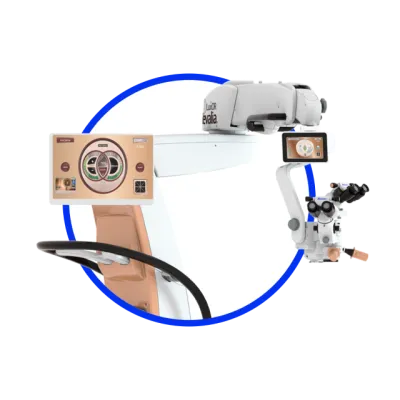
LuxOR® Revalia™ - Ophthalmic Microscope
The LuxOR® Revalia™ Ophthalmic Microscope is designed for superior visualization2,3 and usability for all ophthalmic procedures.
Advanced: Experience unprecedented red reflex stability
ILLUMIN-i technology transforms the way light is delivered into the eye.
On traditional ophthalmic microscopes, the objective lens is positioned below the light source, so light beams focus down to a small point. With Alcon’s proprietary ILLUMIN-i® technology, the objective lens is positioned above the light source, so light beams remain nearly collimated.
This innovative design provides:
- An expanded illumination field with a 6x-larger, highly stable red reflex zone1,2
- Greater red reflex stability during patient eye movement4
- Crisp visualization during every intraoperative phase
- Longer safe exposure time, compared to Zeiss Lumera†, to help minimize the risk of phototoxicity during cataract surgery
LuxOR® Revalia™ features the following unique technologies that are designed to optimize both anterior and posterior segment procedures.
Anterior Segment Procedures
- Expand visibility with a 6x-larger red reflex zone with greater stability than other microscope systems
- A large depth of focus allows you to see more planes of the eye at once
- Experience seamless integration with the ARGOS® Biometer with Image Guidance, which provides digital markers and image-guided overlays throughout your procedure
- Monitor CENTURION® Vision System surgical status on live video overlays via the rear control panel and external display
- Compared to Zeiss Lumera*, longer safe exposure time for reduced risk of phototoxicity† during cataract surgery
- Longer life and customizable settings with LED illumination
Posterior Segment Procedures
- A large depth of focus allows you to see more planes of the eye at once
- High magnification binocular options allow for better visualization during macular procedures
- Q-VUE® Assistant allows for a dedicated 3D stereo assistant view with independent fine focus
- Utilize proprietary AMP technology to shift the beam splitter out of your optical path and brighten the image for the surgeon and tech
- Support your visualization of the retina full functionality of the OCULUS BIOM*, wirelessly operated by the LuxOR® Microscope foot controller
- The LuxOR® screen now dims to allow for a darker vitreoretinal surgical environment