Horiba SWIFT - Ultra Fast Raman Imaging Software
SWIFT ultra-fast Raman imaging allows detailed Raman maps can now be acquired on second/minute timescales with integration times down to 1ms and below. SWIFT retains the true confocal performance of the HORIBA Scientific Raman systems, thus ensuring optimised spatial resolution for analysis of small particles and thin layers.
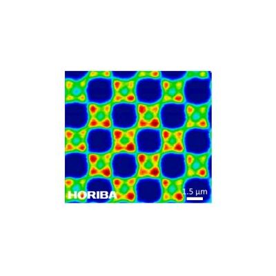
Second generation SWIFT ultra-fast Raman imaging, showing real time hyperspectral Raman image acquisition on a structured semiconductor device, with 40,200 spectra acquired in less than 50 seconds.
Fast Raman imaging offers many advantages to the user:
- Acquisition times reduced by orders of magnitude without compromise in image quality.
- Macro areas (centimeters) can now be analysed with micro resolution.
- High definition megapixel Raman images are now possible in a single fast Raman map, providing both a broad overview and detailed high resolution work in a single experiment.
- Data rich 3D confocal volume images can be acquired in realistic timescales, allowing a sample’s internal structure to be interrogated.
- Time resolved Raman imaging – with a full image acquired in just a few seconds, chemical reactions such as polymer curing occuring on minute/hour timescales can now be imaged.
SWIFT is suitable for use with all lasers from UV through to near infra-red. It is compatible with all HORIBA Scientific systems, either at time of purchase, or as a retro-fittable upgrade (additional or replacement parts may be required).
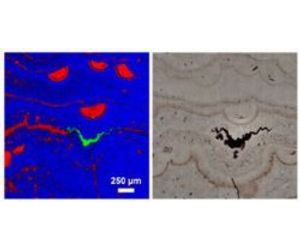
SWIFT ultra fast Raman image of a mineral section, illustrating distribution of quartz species (red/blue) and a haematite seam.
SWIFT Raman image of graphene on Silicon, showing mono-layer, bi-layer and tri-layer areas. A step size of 200nm was used, with 50 ms integration per point.
SWIFT 3D Raman volume display of barium sulfate particles in a polymer matrix in a 92μm3 volume, acquired with 200nm step and 50ms integration.
High definition SWIFT image of entire 17mm pharmaceutical tablet, comprising over 2.6 million spectra. Zoom regions are taken from the same data, illustrating how a single high definition image can be used to survey on millimeter scales and investigate detail on the micrometer scale.
SWIFT ultra-fast Raman imaging of onion cells, illustrating general cell structure (red/blue) and isolated zones of carotenoid species.