

- Home
- Companies
- Meagan Medical, Inc.
- Products
- Meagan - Model CFS 2.0 - Cutaneous ...
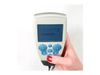
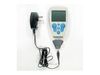
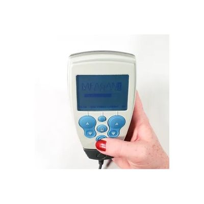
Meagan - Model CFS 2.0 -Cutaneous Field Stimulation Device
Cutaneous Field Stimulation (CFS) is a novel form of electrotherapy to manage pain. CFS can provide 3-6 hours of pain relief following a 30 minute treatment. The action of CFS is achieved by stimulating specific types of nerve fibers, leveraging the nervous system anatomy, to essentially divert the brain’s attention from the signal of chronic pain. The result is a reproducible, longer duration of pain relief. This non-curative therapy is envisioned as a cost effective adjunct to comprehensive pain management program to improve patient quality of life and reduce overall health care costs. It is reasonable to predict that improved pain control will foster increased activity and the associated strengthening which results from increased activity should provide long lasting functional improvement as well.
Cutaneous Field Stimulation (CFS) uses a unique array of tiny needle-like electrodes that are specifically designed to stimulate superficial nerve fibers (cutaneous Ad and C fibers). It uses low frequency (4 Hz), high current density stimulation via needle like electrode arrays that only penetrate the first layer of the epidermis (Stratum Corneium). The electrode arrays (Transducers) are placed directly over the area of pain.
There is a hierarchy in the processing of signals in the spinal cord. Signals from Ad fibers transmit urgent messages about injuries to the body, they alert the brain to act quickly to take evasive action. (Think Ad fibers = Acute pain). Signals from C fibers transmit less urgent messages which signify achey or dull type pain. (Think C fibers = Chronic pain). The spinal cord prioritizes signals from Ad fibers over signals from C fibers. An example would be that if your arm muscles ache from a hard work out the signal is carried by C fibers but if you hit your thumb on that arm with a hammer the acute pain will be carried by Ad fibers and your reaction will be to ignore the achy pain and jerk your arm away even though your arm still aches. As long as there are volleys coming from Ad fibers, the spinal cord will suppress the perception of signals from C fibers. Thirty minutes of asynchronous stimulation of the Ad fibers by CFS has a long-term depressive (LTD) effect on the perception of local C-fiber activity (Chronic pain). This effect occurs in the spinal cord and can provide pain relief for 3 – 6 hours after therapy has stopped.
The human nervous system features a variety of nerve fibers, each carrying a different type of sensation to the spinal cord, which, in turn, forwards messages to the brain. Of particular interest are A beta (Aβ) fibers, A delta (Aδ) fibers, and C fibers. Aβ fibers transmit non-painful mechanical sensations, such as those generated from rubbing and touching. Aδ fibers transmit sharp, intense, immediate pain that signals that injury is taking place and that some action is urgently needed to evade the source of injury. C fibers transmit the dull, aching, long-term pain that is the hallmark of chronic pain syndromes. Both Aβ and Aδ fibers are myelinated, so they transmit signals to the spinal cord very quickly. C fibers are unmyelinated, so their signals travel slowly compared to the Aβ and Aδ fibers.
Conventional TENS works according to gate theory, which posits that activation of the non-nociceptive (non-painful) Aβ fibers interferes with the pain signals coming from C fibers. This interference takes place in the spinal cord. Human beings use the gate-theory mechanism intuitively: for example, when people accidentally hit their elbows (activating the fast pain-transmitting Aδ fibers), they intuitively rub the site of the injury, activating the faster Aβ fibers to reduce the perception of pain.
A lesson learned from gate theory is that signals travelling along fast-conducting nerves will reach the spinal cord before signals traveling along slow-conducting nerves, allowing them to block the transmission of the slower signals to brain. This blocking mechanism reduces the perception of pain. The mechanism of TENS is to activate Aβ fibers to block signals coming from relatively slow C fibers.
Limitations of TENSTENS works best for general pain—pain from healthy nerve fibers reporting injuries in the body to the spinal cord and brain. For this reason, TENS is best for short-term pain. The effects of TENS generally last for the duration of stimulation therapy and for 15 or 20 minutes after therapy is stopped. For chronic pain, it isn’t practical to deliver TENS therapy continuously to prevent the recurrence of pain.
The importance of spatial distributionEven if Aβ fibers are transmitting signals to the spinal column faster than the C fibers carrying chronic pain signals, they might not be reaching the right parts of the spinal cord to block the C fibers. Nerve fibers enter the spinal cord through nerve roots and then synapse (join) branches of nerves above and below the nerve roots. Aβ fibers and C fibers branch up and down the spinal column for approximately the same distance (Figure 1). A portion of the C fibers may have branches that extend above the Aβ fibers. C fibers connecting to the spinal cord above the Aβ fibers activated by TENS will not be blocked by the Aβ fibers, so the spinal cord will transmit their pain signals to the brain.
How CFS is Different
Cutaneous field stimulation (CFS) is a technique for stimulating Aδ fibers, based on the ground-breaking research of Jens Schouenborg, PhD, of Lund University in Sweden.
CFS activates Aδ fibers instead of Aβ fibers. Aδ fibers aren’t as fast as Aβ fibers, but, as myelinated fibers, they are still much faster than the C fibers that transmit chronic pain. Aδ fibers are more effective than Aβ fibers at blocking signals from C fibers, for a number of reasons. First, Aβ fibers and C fibers both connect to the spinal cord at approximately the same depth of the cord (laminae II and III); however, Aδ fibers penetrate the spinal cord across several layers (laminae I through IV), as shown in Figure 2. Furthermore, Aδ fibers cover a wider area of the spinal cord. Whereas Aβ fibers branch up and down the spinal cord approximately the same distance from the nerve root as the problematic C fibers, Aδ fibers branch up and down the spinal cord much further than C fibers. With CFS, it is less likely that branches of C fibers will extend above the activated Aδ fibers, so more of the chronic pain signals are blocked (Figure 3).
In effect, there is a hierarchy in the processing of signals in the spinal cord. The spinal cord prioritizes signals from Aδ fibers over signals from C fibers. Since Aδ signals transmit urgent messages about injuries to the body, they signal the brain to act quickly to take evasive action. As long as there are signals coming from Aδ fibers, the spinal cord will suppress signals from C fibers.
CFS’s 14 electrodes, firing one a time, generate a field of low-level Aδ nerve activation over the area where the patient feels chronic pain (Figure 4). The barrage of Aδ signals travels to the spinal cord, where the wide distribution of Aδ signals covers the narrower distribution of C-fiber signals, and the spinal cord prioritizes the relatively comfortable Aδ signals over those of the painful C-fiber signals.
Furthermore, the asynchronous stimulation of the Aδ fibers by CFS has a long-term depressive (LTD) effect on the perception of local C-fiber activity. This effect occurs in the spinal cord and provides pain relief for 3 –6 hours after therapy has stopped.