

- Home
- Companies
- Imagin Medical
- Products
- Cystoscopy with the i/Blue Imaging ...
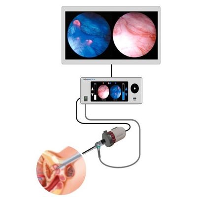
Cystoscopy with the i/Blue Imaging System
Imagin Medical’s i/Blue Imaging System addresses the limitations of both white and blue light cystoscopy while maintaining the advantages of both. By combining innovative optic and light sensor technology with the same FDA-approved imaging agent, surgeon’s will be able to view real-time, side-by-side white and blue light images on the same monitor, eliminating the need for surgeons to switch back and forth. This method has the potential to make bladder cancer detection and removal more efficient and accurate, as well as reduce recurrence rates and health care costs.
Unlike current bladder cancer visualization systems, the i/Blue Imaging System is a device external to the body with the capability to attach to most endoscopes currently on the market. As a result, hospitals will be able to integrate Imagin Medical’s technology with endoscopes they already own. While the current blue light method requires a system tower that houses the light source, camera control and video data recorder units, the i/Blue Imaging System consolidates this instrumentation, combining these three modules into one compact device.
- Makes blue light cystoscopy practical and more accessible to hospitals and patients
- Enhances visualization of cancerous cells for more accurate removal
- Displays simultaneous, side-by-side white and blue light images on the monitor
- Eliminates the need for surgeons to switch back and forth
- Shows cancer in context within the bladder
- Consolidates state-of-the-art light source, camera control and video data recorder into one compact device
- Adapts to most endoscopes on the market, allowing hospitals to use surgical equipment they already own