
Ivantis - Hydrus Microstent
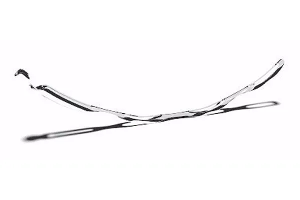
Roughly the size of an eyelash, the highly flexible Hydrus Microstent is placed by a doctor using microscopic incisions and advanced equipment. This less invasive approach allows for fewer complications and faster healing times than traditional glaucoma surgery.
Hydrus Microstent helps to reduce eye pressure in 3 ways
The Hydrus Microstent was designed to enhance fluid outflow in multiple ways to help patients to reduce eye pressure.
Enhancing outflow
Hydrus Microstent is placed in Schlemm’s canal, a part of the drainage system of the eye. Fluid then flows along the canal (via the Hydrus Microstent) and into the eye’s natural outflow channel to reduce eye pressure.*
Expanding the eye’s natural fluid pathway
Hydrus Microstent delicately expands the natural width of Schlemm’s canal, allowing for enhanced flow through the eye’s drainage system to help reduce eye pressure.
Engineered to reduce eye pressure
Hydrus Microstent spans approximately 90° within the eye. This coverage ensures that fluid reaches the eye’s drainage system, which carries fluid from Schlemm’s canal to the body’s circulatory system to reduce eye pressure reliably.
Implantation of the Hydrus Microstent is straightforward and the device is delivered via a preloaded stainless steel cannula. Advantages of the procedure include:
Ab interno canal-based approach
Implantation into Schlemm’s Canal, the eye’s natural drainage channel, which is considered a safe anatomical target and a suitable first-line therapy for implantable MIGS devices
Confirmation of placement
Immediate and direct visual confirmation of successful implantation with minimal learning curve1
No stent targeting
Approximately 90° span ensures access to multiple collector channels, and eliminates the need for stent targeting