NvisionVLE - Imaging System with Real-time Targeting
The NvisionVLE Imaging System, with Real-time Targeting, as part of the VLE procedure (Volumetric Laser Endomicroscopy), uses an optical signal acquisition and processing method to create high-resolution cross-sectional images and mark tissues—letting you evaluate 100% of the tissue in a 6cm scan in real-time. 3mm deep. At a resolution of 7 microns. All so you can provide a more thorough evaluation – potentially leading to improved biopsy targeting for diagnosis by histopathology, and more complete information to determine the best treatment for your patients.
The NvisionVLE Imaging System is indicated for use as an imaging tool in the evaluation of human tissue microstructure, including esophageal tissue microstructure, by providing two-dimensional, cross-sectional, real-time depth visualization and may be used to mark areas of tissue. The safety and effectiveness of this device for diagnostic analysis (i.e. differentiating normal versus specific abnormalities) in any tissue microstructure or specific disease has not been evaluated.
AThe NvisionVLE Imaging System with Real-time Targeting allows clinicians to evaluate the esophageal tissue microstructure and mark areas of suspicion that are not visible with conventional imaging modalities, during a standard endoscopy procedure. These features help provide a more thorough evaluation—potentially leading to improved biopsy targeting for diagnosis and more effective therapeutic decisions for patients.
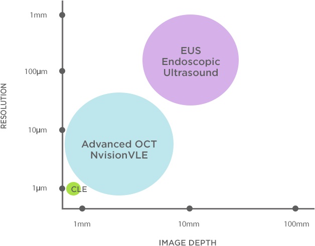
- Uses Advanced OCT to capture images up to 3mm beneath the mucosa at a 7 micron resolution in real time—unlike white light endoscopy, which can only image surface detail
- Offers a volumetric view (~10,000mm2), as opposed to a “point” image typically obtained with confocal microscopy (0.25mm2)
- Advanced Optical Coherence Tomography (OCT) imaging delivers up to 25X higher resolution than endoscopic ultrasound
- Provides a dramatic increase in imaging speed and improved image resolution, compared to first-generation OCT systems
- Creates tissue laser marks visible under white light endoscopy, designed to help clinicians target biopsies at a site of interest that may not be visible with other imaging modalities
Explore the entire targeted segment of the esophagus in real time:
- Collect 1,200 cross-sectional images
- Across a targeted segment of 6cm
- Penetrating approximately 3mm into esophageal tissue
For close examination of a particular area of interest, either within the cross-sectional or longitudinal views, these windows provide a zoomed-in view.
Longitudinal ViewExamine the plane of the esophagus perpendicular to its cross-section:
- View the esophageal wall along the axis of the organ
- With over 4,000 longitudinal images of the esophagus
As you manipulate the cross-sectional or longitudinal views on the NvisionVLE touch-screen monitor or hand-controller, each of the corresponding views update smoothly and in real time.
- Hand-controller allows for real-time physician control of the VLE scan and tissue marking to better evaluate areas of concern
- Active clinican-controlled workflow, tailored to the specific needs of each patient
- Single and double mark options for application flexibility
- Balloon catheter facilitates optical probe positioning and centering (available in 14mm, 17mm and 20mm sizes)
- Also available as balloon-less 7 French Low-Profile optical probe to accommodate various anatomies
- Catheter compatible with endoscope channels 2.8mm or larger
The Current Procedural Terminology (CPT)* codes used for reimbursement of the VLE (Volumetric Laser Endomicroscopy) procedure are:
- CPT 43206 Esophagoscopy, flexible, transoral; optical endomicroscopy
- CPT 43252 Esophagogastroduodenoscopy, flexible, transoral; optical endomicroscopy
- CPT 0397T – Endoscopic retrograde cholangiopancreatography with optical endomicroscopy
- CPT 88375 Optical endomicroscopic image(s), interpretation and report, real-time or referred, each endoscopic session
A Faster, Better Way of Seeing
NinePoint Medical’s NvisionVLE® Imaging System with Real-time Targeting&trade is a groundbreaking convergence of superior optical imaging, innovative engineering techniques, and revolutionary information technology. At the heart of the system, Advanced Optical Coherence Tomography (OCT) technology provides physicians and pathologists with comprehensive, volumetric digital images of a patient’s esophagus. This next-generation technique enables much higher imaging speeds, combined with improved field-of-view and signal-to-noise ratio.
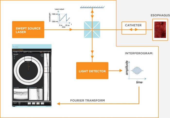
The (greatly simplified) fundamentals of Advanced OCT are shown in the diagram. Similar to a traditional laser, the swept-source laser produces light of only one ‘color’ at any instant in time. However, unlike a traditional laser, the swept-source changes the color of its output light very rapidly in time.
The output light is split in two: one beam stays internal to the console, while the second is transmitted through the catheter and focused into the tissue. Some of the light is reflected back from physiological structures within the tissue, and this reflected light is collected by the catheter and returned to the console.
Inside the console, the reflected and internal beams are combined and interfere with each other. After some processing, the interference signal yields an axial line (‘A-line’) that records the intensity of reflections at each depth in the tissue. Combining many consecutive A-lines into an image provides a cross-sectional look at all the reflecting structures in the tissue.
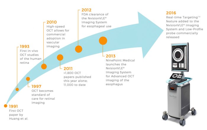
The basic principles of Optical Coherence Tomography (OCT) were initially used within the fields of optical radar and optical telecommunications, among others. The technology expanded in the late 1980s when researchers began exploring applications for biological tissue imaging. Over the next decade, researchers around the world developed sub-categories of the technology, such as swept-source OCT, and began investigating ways to use OCT in different types of tissues and biomedical applications.
Today, commercial OCT systems are used regularly as an imaging standard in ophthalmology, cardiology, dermatology, and general research. NinePoint’s Advanced OCT technology, licensed from the Wellman Center for Photomedicine at Massachusetts General Hospital, initially focuses on esophageal and general imaging of tissue microstructure.