

EnFocus - Intraoperative OCT Imaging System
Apply your skills with even greater confidence during complex anterior and posterior segment surgeries with a Leica OCT microscope solution.
EnFocus intraoperative optical coherence tomography (OCT) allows you to see what lies underneath the surface. It gives you the additional real-time information you need for a deeper understanding of how subsurface tissue reacts to your surgical maneuvers.
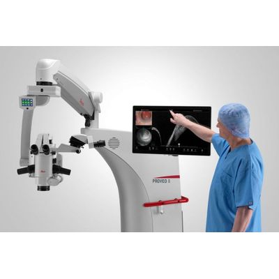
Our intraoperative OCT imaging system is available fully built into the Proveo 8 ophthalmic microscope. You can enhance the microscope view with OCT imaging at any step during surgery with just a few taps. The Proveo 8 microscope with integrated EnFocus OCT supports you to focus on achieving an optimal patient outcome by providing:
- Greater insight into hidden subsurface details
- Immediate confirmation on how tissue reacts to your surgical maneuvers
- Maximum freedom in viewing and reviewing optimized OCT images
Greater Insight: See more details below the surface
Supplement your microscope view with bright, sharp imaging of previously hidden subsurface tissue details. Intraoperative integration of OCT imaging provides you with additional information so you can get greater insights into ocular pathology during surgery.
- Clearly differentiate between artifacts and tissue due to the unique spectrometer technology with dispersion compensation software and a highly sensitive detector that captures more signal
- See fine details even through blood in trauma cases thanks to an axial resolution of 2.4 μm in tissue
- Capture comprehensive area scans with high lateral resolution, due to a high scan density of up to 1000 A-scans x 1000 B-scans to not miss out on an important detail
- Playback through acquired OCT scans frame by frame or in video mode to check e.g. if there is residual sub-retinal fluid, if the positioning of the glaucoma drainage device is correct, or if the corneal graft is well opposed to the host cornea.
Greater insight: Get a wider view
Ensure you have a full view of the surgical field so you don’t miss features at the periphery. EnFocus provides a 20 x 20 mm lateral field of view even at high magnification.
No need to adjust the viewing perspective during surgery. This enables you to monitor any changes at the periphery even when working in the center of the eye.
EnFocus offers wide scans, that cover the whole cornea and the anterior chamber – detail and resolution are impressive and allow a thorough visualization of anterior segment details. EnFocus provides a remarkable insight into the third dimension, meaning the vertical axis Z.
Enrico Bertelli MD, Head of Ophthalmic Department, Bolzano Hospital, Italy.
Greater insight: Clinical assessment to intraoperative decisions
Bridge the gap between pre-operative assessment and real-time evaluation of changes to tissue microstructures during surgery with EnFocus OCT.
In a clinical setting, OCT has become the standard of care with clinical OCT imaging such as Envisu C2300. OCT imaging systems are now moving more and more into the operating rooms where they are impacting surgical decision-making. The DISCOVER Study* collected data on the impact of intraoperative integration of OCT . The study showed that microscopes with OCT provide additional information and can consequently change the surgical plan intraoperatively. In membrane peeling procedures the study showed that intraoperative OCT findings were discordant from the surgeon`s initial impression in 35% cases.
Benefits for your retina surgery
Use OCT to assess the level of tension in a membrane peel in real time, in order to avoid potential tears and protect the integrity of underlying tissue. A high-resolution view of <=4 µm also aids examination of retinal morphology for residual membranes and complications such as macular holes, or sub-retinal edema.
Additional benefits for posterior surgery include:
- Dynamic scan control via footswitch for swift adjustment of the scan angle to align with the membrane tissue
- Easy integration of fundus viewing systems such as the BIOM5 from OCULUS with synchronized focus
Immediate confirmation: Observe & respond to tissue changes
The EnFocus intraoperative OCT imaging system provides real-time confirmation of how ocular tissue is reacting to your surgical maneuvers. You can immediately adjust your surgical plan if needed for greater confidence in the surgical outcome.
Observe real-time tissue reaction and respond instantly
- Real-time display of 30 fps provides immediate feedback at each step e.g. to verify adherence of donor tissue in DMEK or DSAEK surgery
- If OCT reveals a complication which wasn’t visible via the microscope view, for example due to bleeding, you can instantly adapt your surgical plan
- For additional verification you can easily review or playback through the acquired scans frame by frame or in video mode
- Live on-screen measurements provide even more information e.g. cornea thickness and needle depth during DALK surgeries
Maximum Freedom: Smooth independent workflow
EnFocus built into the Proveo 8 supports your workflow and enables you to independently control your OCT view. You are free to concentrate on your procedure.
- Easily activate intraoperative OCT yourself at any point during surgery via footswitch, handle, or 27” touchscreen HD monitor
- Preprogram your personal settings and modes before surgery according to surgery type and step then allocate to the footswitch or handle for a smooth workflow
- Record or capture images in the same way, choose the Evolution4K recording system from Med X Change for comprehensive documentation
- View your microscope and intraoperative OCT-image in the eye pieces via the DI C800 or on the 27” HD touch-screen monitor and for even larger screen projections, four video outputs are available