
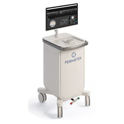
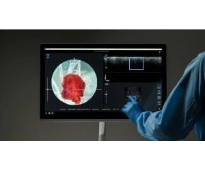
Perimeter - Model S-Series -Optical Coherence Tomography (OCT) System
Harnessing the power of advanced technology and intuitive functionality, Perimeter Medical Imaging is driven to transform cancer surgery with advanced imaging tools. The Perimeter Optical Coherence Tomography (OCT) system provides cross-sectional, real-time margin visualization of tissue microstructures during surgical procedures. Perimeter’s game-changing technology gives surgeons unmatched clarity and confidence on margins at the point of care.
High-resolution OCT
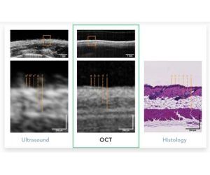
Using light, images have 10x higher resolution than ultrasound and X-ray and 100x higher than MRI for visualization at the cellular level.
Margin Visualization
Resolution is optimized for visualizing microscopic tissue structures down to 2mm, ideal for reviewing surgical margins.
Non-Invasive and Non-Destructive
Tissue is entirely preserved for histopathology and device does not come in contact with the patient or enter the sterile field.
Advanced Software
Identify and annotate regions of interest with image review manipulation tools for optimized decision-making in the operating room.
Orientation Labeling
Label and capture images of surgical margins on all six planes to convey accurate orientation for pathology.
A Powerful Tool for Margin Visualization
High-resolution intraoperative imaging technology
The Perimeter S-Series OCT is designed to provide surgeons with the clinical support they need in the OR — every step of the way. Clinical guidelines state that 2mm of clear margins produce the best outcomes for patients undergoing tissue excision assessment.
Perimeter’s advanced technology delivers the clearest 2mm subsurface imagery available, with 10x the image resolution of standard X-ray and ultrasound and 100x greater than MRI, for intraoperative imaging power that’s truly next level.
Imaging power designed for clinical support right in the OR
Optical Coherence Tomography (OCT)
Using light, images have 10x higher resolution than ultrasound and X-ray and 100x higher than MRI for visualization at the cellular level.
Margin Visualization
Resolution is optimized for visualizing microscopic tissue structures down to 2mm, ideal for reviewing surgical margins.
Non-Invasive and Non-Destructive
Tissue is entirely preserved for histopathology and device does not come in contact with the patient or enter the sterile field.
Advanced Software
Identify and annotate regions of interest with image review manipulation tools for optimized decision-making in the operating room.
Orientation Labeling
Label and capture images of surgical margins on all six planes to convey accurate orientation for pathology.
Surgeons have been asking for a new generation of imaging technology that provides clear, ultra-high-definition imagery during surgery. Perimeter’s S-Series OCT delivers the clarity needed at the point of care — when it matters most.
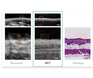
These images show the correlation of the Perimeter OCT volume to histology. This technology does not replace the need for histology, but provides real-time insight on margins for surgeons before closing.
It’s clear to see that the OCT images show cellular-level detail surgeons have never seen before, and it happens all in real time right in the OR.
BREAST
Representative correlation. Top panel: OCT image showing breast tissue microstructures. Bottom panel: H&E slide from corresponding region. (D) Ductal Carcinoma In-Situ (DCIS), (A) Adipose Tissue.
HEAD & NECK
Representative correlation. Top panel: OCT image of the posterior-inferior margin. Bottom panel: H&E slide from corresponding region. (SE) Squamous Epithelium, (S) Submucosal, (SM) Skeletal Muscle, (DLC) Dilated Lymphatic Channel.
THYROID
Representative correlation. Top panel: OCT image showing thyroid tissue microstructures. Bottom panel: H&E slide from corresponding region. (V) Vessel, (F) Follicles.
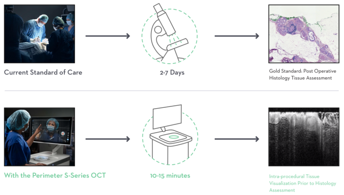
Perimeter S-Series OCT fits seamlessly into your existing workflow. The device itself is portable, so it can be moved into any OR that’s being used for a tissue excision procedure. Scan times are 1-2 minutes per margin or 10-15 minutes overall with interpretation. While it does not replace standard histopathology, OCT provides valuable insights when and where surgeons need to make informed decisions.
Surgeons have worked for years to innovate every step in the diagnosis and treatment plan for their cancer patients, with the ultimate goal of improving survivability and quality of life. Yet, intraoperative margin assessment remains one of their most pressing problems. Watch the clips below to see how one surgeon has incorporated OCT technology into her OR workflow — and now has the tools to visualize margins with ultra-high-definition imagery.
Perimeter’s OCT technology fits seamlessly into the workflow as an adjunct to specimen X-ray and pathology, and it’s portable so it can be easily moved into any OR.
Surgeons can label and capture images of surgical margins on all six planes, with a scan time of 1-2 minutes per margin, to convey accurate orientation for pathology.
Ultra-clear subsurface imagery and image review manipulation tools make it easy for surgeons to identify regions of interest for optimized decision-making in the operating room.
Tissue is entirely preserved and the device does not touch the patient or enter the sterile field. While OCT does not replace histopathology, it can provide guidance for the pathology team.
A high-resolution imaging technology
OCT is a non-invasive optical imaging technique that produces images of subsurface tissue structures. It is similar to ultrasound but uses light instead of sound, resulting in 10x higher resolution. As such, OCT is a powerful tool for visualizing blood vessels, ducts, glands, and surrounding structures. OCT was developed at MIT in the 1990s, and has been widely used in clinical settings ranging from ophthalmology (retina) and interventional cardiology (vessel plaques), to dermatology (skin lesions). It is now being used for new applications, including visualization of excised tissue and shaved margins.
- High-resolution visualization of tissue at cellular level
- Direct correspondence to histological appearance of tissue structures
- Incredibly fast and portable, allowing for intraoperative tissue visualization
- Two- and three-dimensional image generation
Light interactions reveal different tissue characteristics
A single beam of light is directed at the tissue specimen and rapidly moved/translated across the desired scan area. Light that is reflected back from the tissue specimen down from a depth of 2mm is transformed into an OCT image.
REFLECTION
- Very dense features will reflect the light completely:
- Calci?cations
- Surgical clips, wires
TRANSMISSION
- Less dense features allow light to pass through them:
- Adipose tissue
- Cysts
SCATTERING
- Denser features cause the light to scatter:
- Fibrous tissue
Each light beam captures a single depth profile of a specific area. This single image is called an axial A-scan. As the beam of light moves across the tissue in a line, it generates a long sequence of A-scans that can be compiled into a two-dimensional image known as a B-scan. A series of B-scans can then be stacked to form a three-dimensional image volume.