

- Home
- Companies
- Imedos Health GmbH
- Products
- Imedos - Static Vessel Analysis
Imedos - Static Vessel Analysis
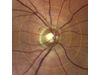
- The imaging system automatically sends a standardised image of the ocular fundus to the software.
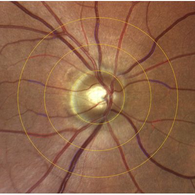
- In this image all essential arterial and venous vessels will be automatically marked according to the protocol of the ARIC study. A measurement grid (ring zone) is placed on the image.
- The SVA system now determines the static vessel parameters for these areas including:
- Central retinal arteriolar equivalent (CRAE): arterial model vessel diameter
- Central retinal venular equivalent (CRVE): venous model vessel diameter
- Arteriolar-to-venular ratio (AVR): CRAE/CRVE ratio
These parameters are valid biomarkers that can be used as risk factors or prognosis indicators for vascular diseases and vascular events in the eye as well as other organs.
Detecting risk factors early on: Fast and precise assessment of the vascular condition using the systems for static vessel analysis
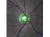
Our product portfolio includes various systems for static vessel analysis. Varying in price, the individual solutions combine an imaging system with the innovative analysis software VesselMap aric and offer different options, such as various image angles or the possibility of fluorescence angiography. Furthermore, they can easily be integrated into already existing practice environments.
You can also update your existing imaging systems. Our Customer Service is happy to advise you and check if your existing hardware is compatible with our products.
The software is not a medical device according to the Medical Device Regulation (MDR) and the Medical Device Law Implementation Act (MPDG), but is intended for research use.
Simple, programme-guided application
Automatic and precise determination of vascular parameters
Suitable as screening method and for use in individualized medicine
Integrated function for follow-up examinations to significantly improve reproducibility and fully automatic evaluation of subsequent images
Patient-related storage of results
When used in combination with a non-mydriatic imaging system, no dilation of the pupil is required for the examination.
The analysis software VesselMap aric can also be used with existing imaging systems (fundus camera) and hardware components. For this purpose, the analysis and calculation parameters of the software are adapted to your system and adjusted accordingly. Individual solutions offer you additional benefits, including:
Protocol functions individually tailored to your practice
Doctor-specific patient database for analysis results
Direct network integration
Image transfer with different image standards such as Dicom
In order to combine the software with your existing imaging system, the following hardware requirements must be met:
Suitable fundus camera
Laptop or PC (Windows 10/11)