
- Home
- Articles
- canada northwest territories
- tissue imaging
Refine by
Tissue Imaging Articles & Analysis
8 articles found

Histological image analysis has emerged as a pivotal domain in biomedical research and clinical diagnostics, allowing scientists and healthcare professionals to decipher the intricate structures of tissues at a microscopic level. This analysis encompasses the examination of biological tissues to understand their composition, organization, and pathology, which can be crucial for diagnosing ...
Dendrimers are highly branched polymers with a three-dimensional structure. They are characterized by expansive functional groups on their surface, which can be tailored to interact with specific molecules or to be used in a variety of applications. Dendrimers have attracted much attention in drug delivery, imaging, and materials science due to their unique structure, high monodispersity, and ...

The use of Quantitative Susceptibility Mapping (QSM) is often viewed as incompatible with current standard radiology workflows. Some radiologists or departments may be unaware of its existence or purpose. Despite the skepticism surrounding its viability in a clinical setting, QSM shows great potential in a wide range of clinical applications, particularly for biomarker detection and ...

Since the 19th century, the practice of tissue processing has remained largely unchanged, resulting in solidified, paraffin-embedded tissue blocks for sectioning. As staining procedures have advanced and immunohistochemistry is becoming more widely used, high-quality tissue sections are paramount for accurate interpretation of disease ...

Abstract Cellular function in tissue is dependent on the local environment, requiring new methods for spatial mapping of biomolecules and cells in the tissue context1. The emergence of spatial transcriptomics has enabled genome-scale gene expression mapping2,3,4,5, but the ability to capture spatial epigenetic information of tissue at the cellular level and genome scale is lacking. Here we ...
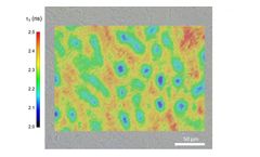
Welcome to Edinburgh Instruments newest blog celebrating our work in Raman, Photoluminescence, and Fluorescence Lifetime Imaging. Every month we will highlight our pick for Map of the Month to show how our spectrometers can be used to reveal all the hidden secrets in your samples. April Studying animal tissue is an important tool for scientists to learn more about the effects of treatments for ...
Highlights Deterministic barcoding in tissue enables NGS-based spatial multi-omics mapping DBiT-seq identified spatial patterning of major tissue types in mouse embryos Revealed retinal pigmented epithelium and microvascular endothelium at cellular level Direct integration with scRNA-seq data allows for rapid cell type identification Summary We present ...

Quantum dots have the potential to be used as contrast agents for molecular imaging in vivo, but there are many challenges in optimising such procedures, both in pre-clinical animal models and in the potential clinical applications. In particular, it is critical to obtain the optimal target-to-background contrast to achieve maximum diagnostic accuracy. However, available data are insufficient to ...