

OrganoReady - Blood Vessel HUVEC
OrganoReady® Blood Vessel HUVEC comprises 38 ready-to-use HUVEC tubules for drug exposure, transport, and permeability studies. The product is based on the OrganoPlate® 3-lane 40 with 3 adjacent channels per chip, including one perfused HUVEC tubule. Do you want to perform experiments in human-relevant, complex 3D tissue models but don’t have the time or experience to build your own? Look no further, with the OrganoReady Blood Vessel HUVEC you can now take your vascular research to the next level. Perform your self-created experiments to investigate permeability, absorption and transport, toxicity, and barrier integrity (TEER) in a physiologically relevant model.
- OrganoPlate® 3-lane 40, including:
- 38 chips with ready-to-use HUVEC tubules
- 2 control chips without cells
- Direct access to the apical and basal lumen of tubules
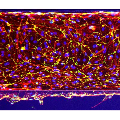
- HUVEC cell line: Human umbilical vein endothelial cells
- ECM: Collagen I (Rat Tail)
Each tissue culture chip contains one in-gel culture channel and two perfusion channels, of which one perfused HUVEC tubule. Compounds and stimuli can be added directly from the apical and basolateral sides of the culture. With this direct access, the platform enables perfusion and supports various barrier function and transport assays.
Meet the OrganoReady Blood Vessel HUVEC
Ready-to-assay blood vessel tubules
38 ready-to-use HUVEC tubules, seeded against collagen I, allowing you to perform assays right away.
We create the model, so you can focus on your assays
Preparation and QC of OrganoReady Blood vessel HUVEC are done by our experts. Use the OrganoReady Blood vessel HUVEC directly after arrival for up to 7 days.
Get inspired by a wide range of possible applications
Study for example (drug-induced) toxicity, transport, use it for disease modeling or for fundamental research on the vasculature.
Apical and basal access
Perfused HUVEC tubules, allowing barrier integrity (TEER), transporter (e.g. P-gp), and permeability assays.
No artificial membranes for free migration, invasion, and exchanges of factors
Unique PhaseGuide™ technology allows cells to interact and migrate freely between channels.
In this video, we show an example of the blood vessel tubules grown against an extracellular matrix (ECM). To assess the tightness of the monolayer we performed a barrier integrity assay.
For this assay, we replace the medium with a medium containing a fluorescent dye. Subsequently, we monitor the leakage of the dye from the top channel, which represents the lumen of the tubule, into the adjacent ECM gel. In the first two columns, leaky tubes were cultured, in contrast to the leak-tight tubes in the rest of the plate.