

Therawis - Model ExosomeDx -Smallest type of Extracellular Microvesicles (EMV)
Exosomes (30-150 nm) are the smallest type of extracellular microvesicles (EMV), produced through inward budding multivesicular bodies (MVB), resulting in intra-luminal vesicles (ILV). ILVs are released into the extracellular media by exocytosis. Exosome-associated proteins are used to isolated microvesicles from biological fluids such as blood, urine or cerebral fluid and to identify the source of their origin. Example of such proteins are tetraspanins (CD9, CD63, CD81), immune regulation molecules (HLA-G, MHCI/II), membrane transport and fusion proteins (Rab5). Secretion of exosomes occurs by almost any “normal cell” such as thrombocytes, cells of the immune system, epithelial cells, or disease tissue, like tumor cells and virus-infected cells.
The molecular composition of exosomes is assumed to reflect (patho-) physiological changes in their cells or tissues of origin, carry DNA, RNA, microRNA, proteins as well as lipids and, therefore, could serve as “liquid biopsy”.
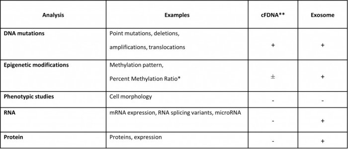
Important cornerstones of cancer treatment, today, are targeted drugs, which are applied once it has been demonstrated that the respective target of the therapeutic is expressed or a specific genetic mutation is present in the cancerous tissue. Tissue is not always available and tissue biopsies are associated with a significant invasive and unpleasant procedure and, therefore, often challenging or impossible to perform, especially in cancer types such as lung or brain tumors and in metastatic disease. Therefore, the blood-based diagnostic approach, the so-called „liquid biopsy“, could solve these issues. Today, the purification of circulating free DNA (cfDNA) and subsequent mutation analysis is used by various in vitro diagnostics such as PIK3CA mutation analysis. cfDNA is able only to address the determination of point mutations, DNA deletions, amplifications and translocations. RNA- and protein analysis cannot be addressed through cfDNA. Exosomes, which supposingly represent the phenotype of the cell of origin, can offer these analyses. The following table highlights the differences in applications of cfDNA vs. exosome analysis.
Exosomes should mirror the phenotype of the parent cell, e.g. from the cell they were released. Exosomes secreted from virus-infected cells, called virosomes, have the potential not only to serve to diagnose the virus type but also the cell population or organ which the virus has infected. Similarly, oncosomes, exosomes released from cancer cells, also should mirror the phenotype of the parental cancer cells. Therefore, identification of oncosomes and characterization of the composition could serve as liquid biopsy for cancer therapy, diagnosis and monitoring.