

Caas - Version MR -Tissue Characterization Software
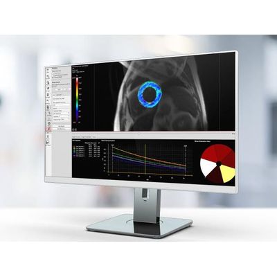
Caas MR Tissue Characterization includes three workflows: Viability, First-Pass Perfusion, and Tissue Mapping. Differentiate between viable and non-viable tissue using regional infarct classification based on delayed-enhanced MR images. Assess myocardial edema based on T2-weighted images. By combining segmental infarct and edema areas, salvageable areas in the area at risk can be identified. Analyze rest and stress perfusion image sets side by side in the first-pass perfusion workflow to determine myocardial perfusion. Tissue mapping by discriminating between T1 and T2 tissue contrast is a unique strength of MRI. It allows the analysis of T1, T2, and T2* relaxation values. Relaxation values are translated into a color map for easy visualization of affected tissue. Visualization of T1 relaxation values is supported for look-locker and modified look-locker sequences.
- Infarct volume and transmurality
- Salvageable area index
- Time-Intensity curve parameters
- Myocardial Perfusion Reserve Index (MPRi) derived by comparing rest and stress
- T1, T2, T2* relaxation values
- Parametric color maps
The Viability Analysis workflow is not 510(k) cleared and therefore not meant for clinical decision making.
- Automatic infarct detection
- Breathing motion correction
- All result types are supported by the AHA 17-segment model