

Pie Medical Imaging B.V. software
Cardiac Diagnostics - Caas MR Solutions
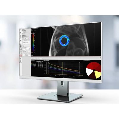
Caas - Model MR 4D - Complete Flow Assessment Software
Caas MR 4D Flow is specifically designed to extract relevant information from 3D phase-contrast (PC) MR images, within a few clicks. Based these phase-contrast images, two pioneering 4D Flow modules are offered to perform a complete flow analysis that can aid in reading and interpreting cardiovascular MR images and assist in planning the time and type of an intervention. These two modules include a module to analyze vessels, from the aorta up to smaller vessels in for example the kidneys (Caas MR 4D Artery), and a module to analyze the heart valves in terms of regurgitation fraction (Caas MR 4D Heart).
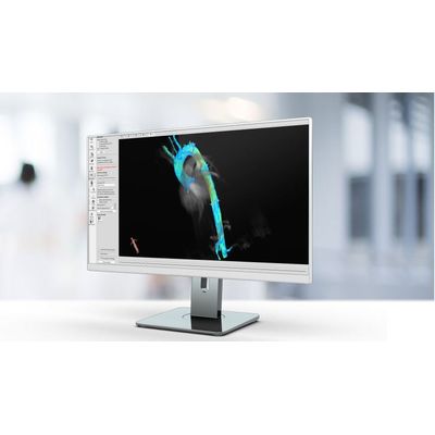
Caas - Functional MRI Images Analysis Software
Designed for the imaging specialist, Caas MR Functional Analysis assists in functional analysis of the heart. The endocardial and epicardial wall of the ventricle can be automatically segmented on short axis images. Long axis images can also be used for segmentation. Ejection fraction, End Diastolic (ED), and End Systolic (ES) volumes are accurately calculated. Wall results in terms of wall motion, wall thickness, and wall thickening can also be obtained.
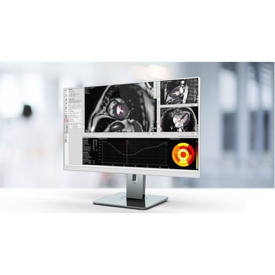
Caas - Model MR - Tissue Characterization Software
Caas MR Tissue Characterization includes three workflows: Viability, First-Pass Perfusion, and Tissue Mapping. Differentiate between viable and non-viable tissue using regional infarct classification based on delayed-enhanced MR images. Assess myocardial edema based on T2-weighted images. By combining segmental infarct and edema areas, salvageable areas in the area at risk can be identified. Analyze rest and stress perfusion image sets side by side in the first-pass perfusion workflow to determine myocardial perfusion. Tissue mapping by discriminating between T1 and T2 tissue contrast is a unique strength of MRI. It allows the analysis of T1, T2, and T2* relaxation values. Relaxation values are translated into a color map for easy visualization of affected tissue. Visualization of T1 relaxation values is supported for look-locker and modified look-locker sequences.
3 Mensio Structural Heart
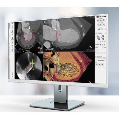
3mensio - Aortic Valve Replacement Software
3mensio Aortic Valve allows you to quickly and reliably pre-plan aortic valve replacement procedures (TAVR/TAVI). The workflow consists of several modules for the assessment and sizing of the aortic root and approach route assessment. Assess for each patient if the transfemoral, transsubclavian, transapical or direct aortic approach is most suitable. The software has an intuitive workflow assistant which acts as a guide through the software making it intuitive and easy to use.
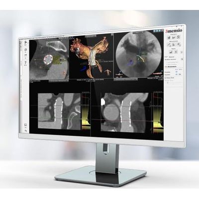
3mensio - Mitral Valve Regurgitation Software
The Mitral Valve is a complex 3D structure. Mitral regurgitation can be treated by replacing the native valve (TMVR) or by repairing the native valve (TMVr). Planning for both types of procedures can be done using the 3mensio Mitral Valve workflow. With a single click we orientate on the mitral annulus. We can do an assisted mitral annulus trace to understand the 3D shape and dimensions of the annulus. Additional features have been developed specifically to plan for replacement or repair. Relationships with surrounding structures like the Aortic Valve and Coronary Vessels can quickly be assessed. An easy comparison between the ED and ES phase can be performed. The approach route can be assessed using either the Transseptal or Direct Access approach module.
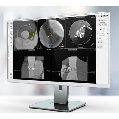
3mensio - Percutaneous Pulmonary Valve Implantation (PPVI) Planning Software
Congenital Heart Disease is a defect in the heart that is present at birth. In a group of these patients the pulmonary valve does not function properly and the valve needs to be replaced. As a prosthetic valve only has a limited lifespan these valve needs to be replaced a number of times during a life. To make the procedure less invasive Percutaneous Pulmonary Valve Implantation (PPVI) has been developed. 3mensio Pulmonary Valve is a dedicated workflow to plan PPVI. The automatic segmentation gives a quick overview of the patient’s anatomy. Different features enable the assessment of the surrounding structures and the planning of a stent and valve.
Vascular Surgery
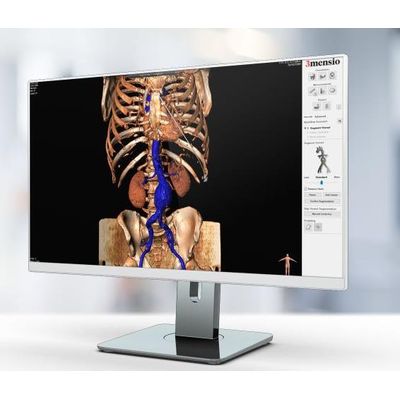
3mensio - Vascular Surgery Software
Easily determine the appropriate landing zone for treatment of abdominal aneurysms (EVAR), thoracic aneurysms (TEVAR) or placement of fenestrated stents (FEVAR). All relevant measurements (e.g. diameter, clock position, volumes and length) can be obtained via automatic detected centerline or 3D double oblique. Integrated manufacturer stent order sheets generate PDF files so specific stents can be easily ordered via e-mail.